
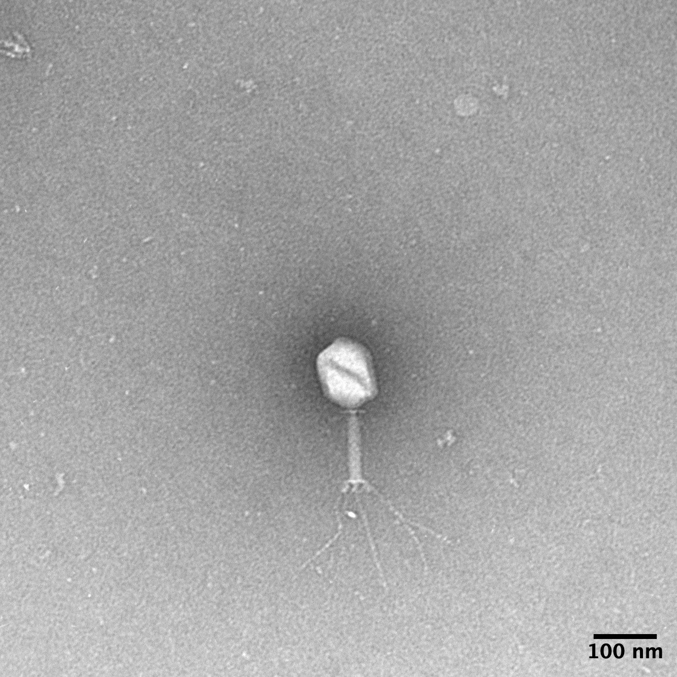
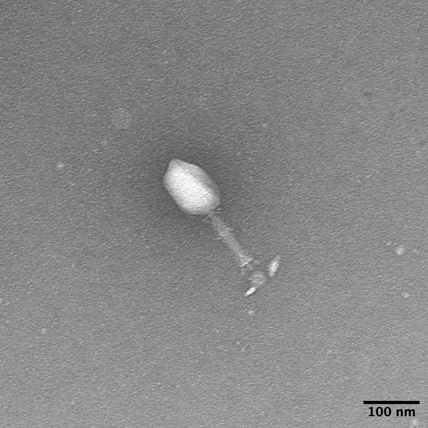
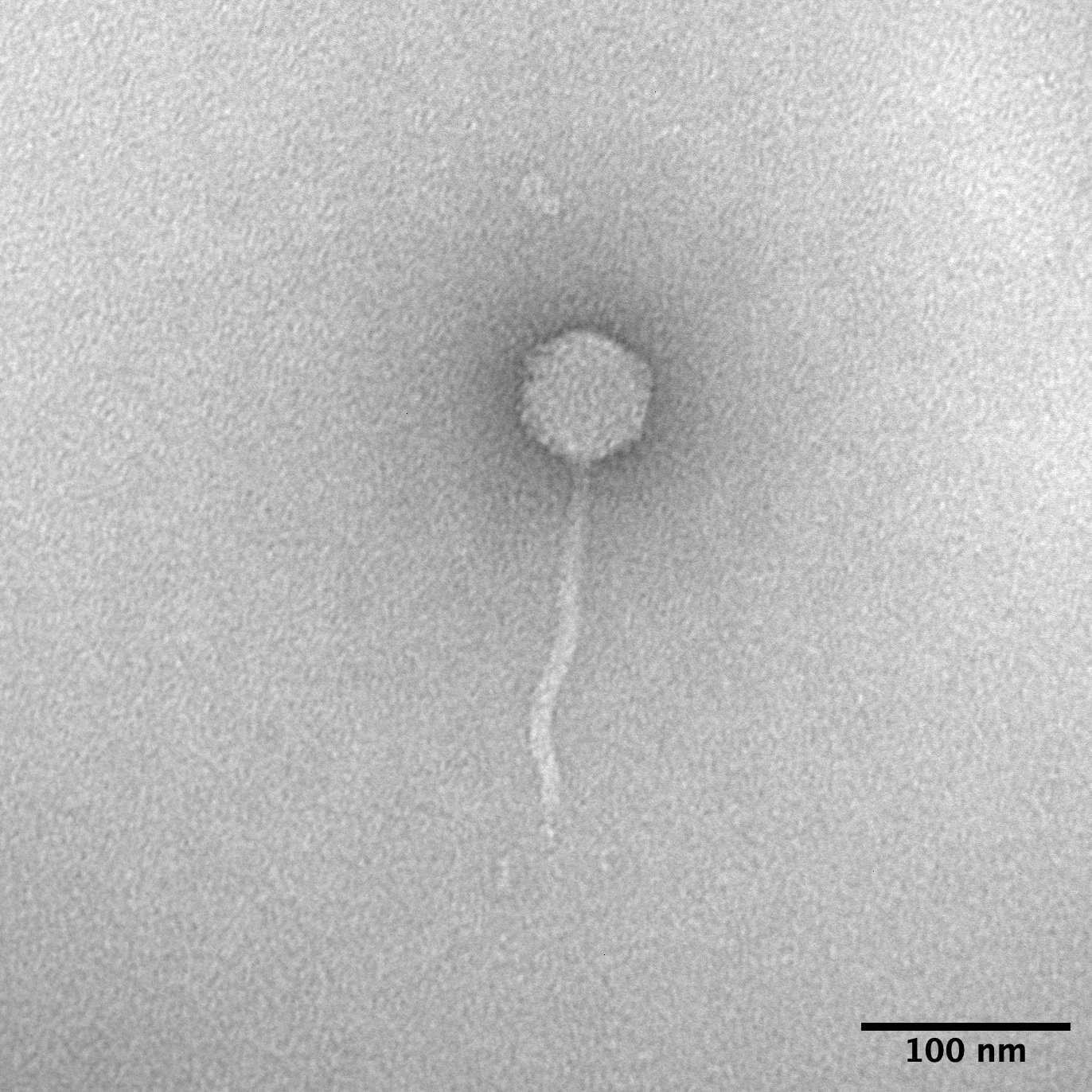
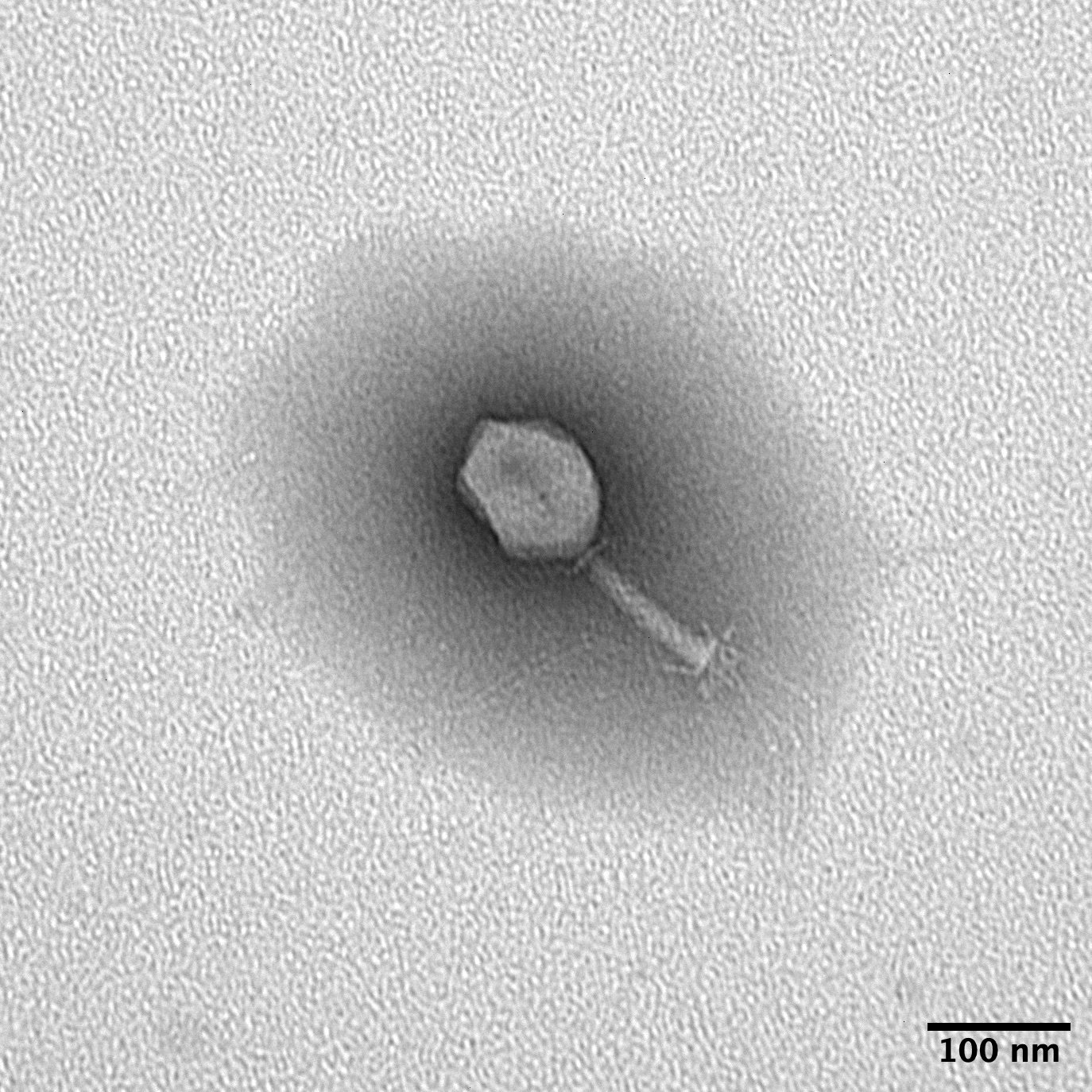
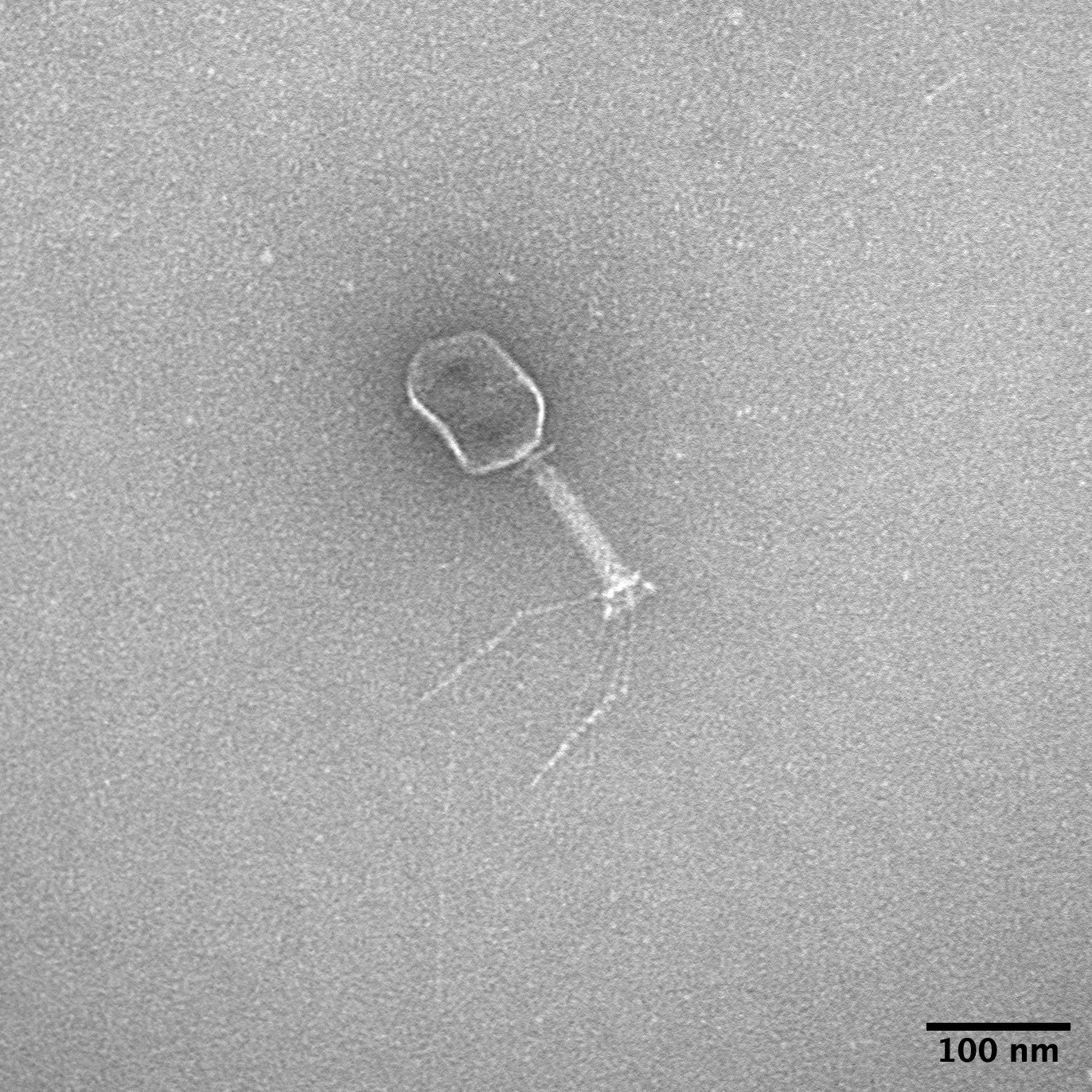
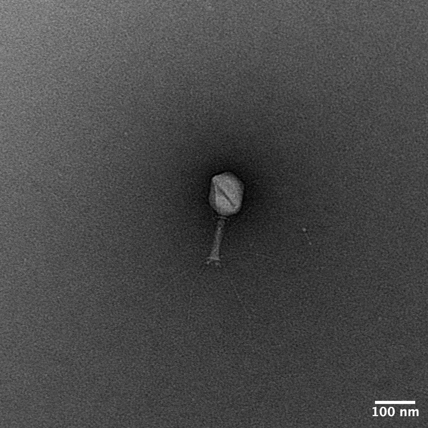
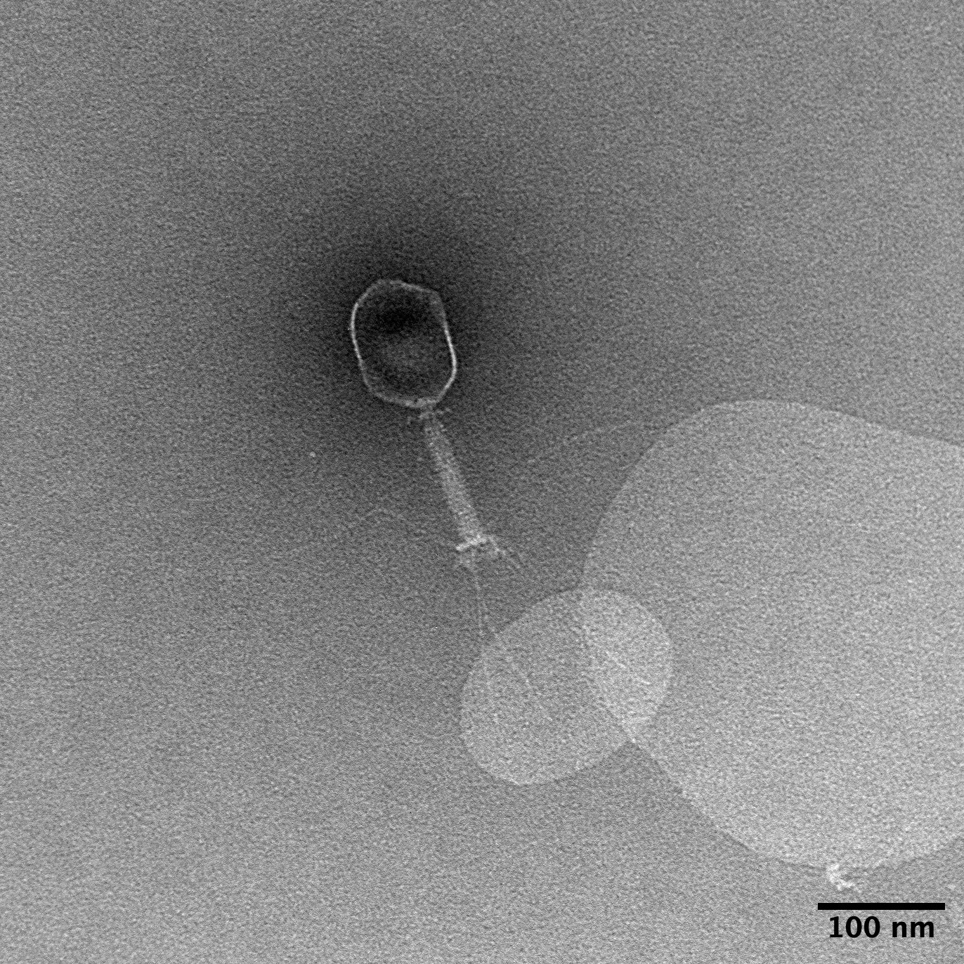
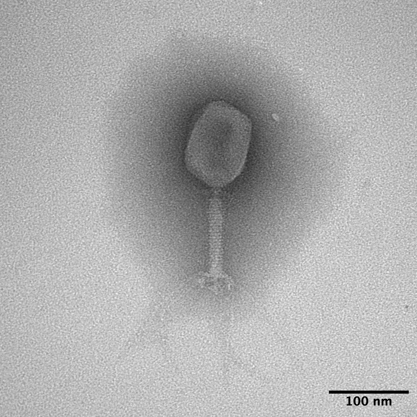
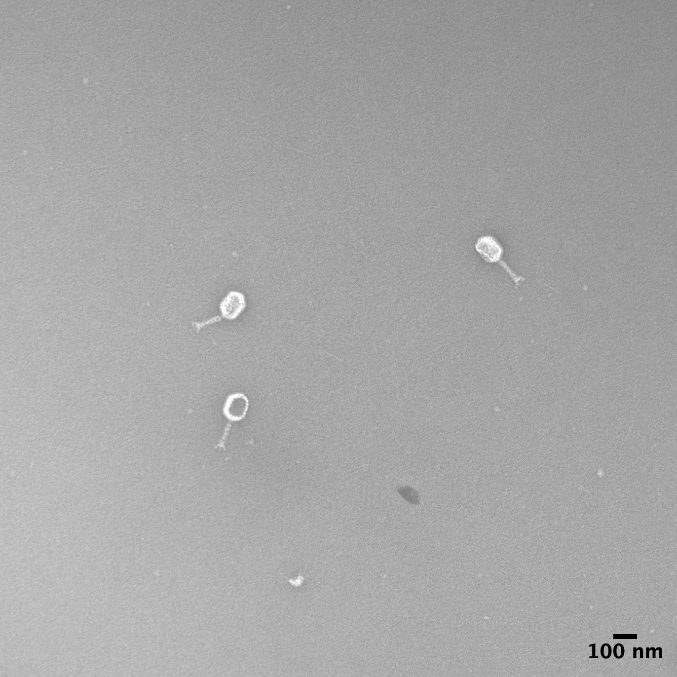
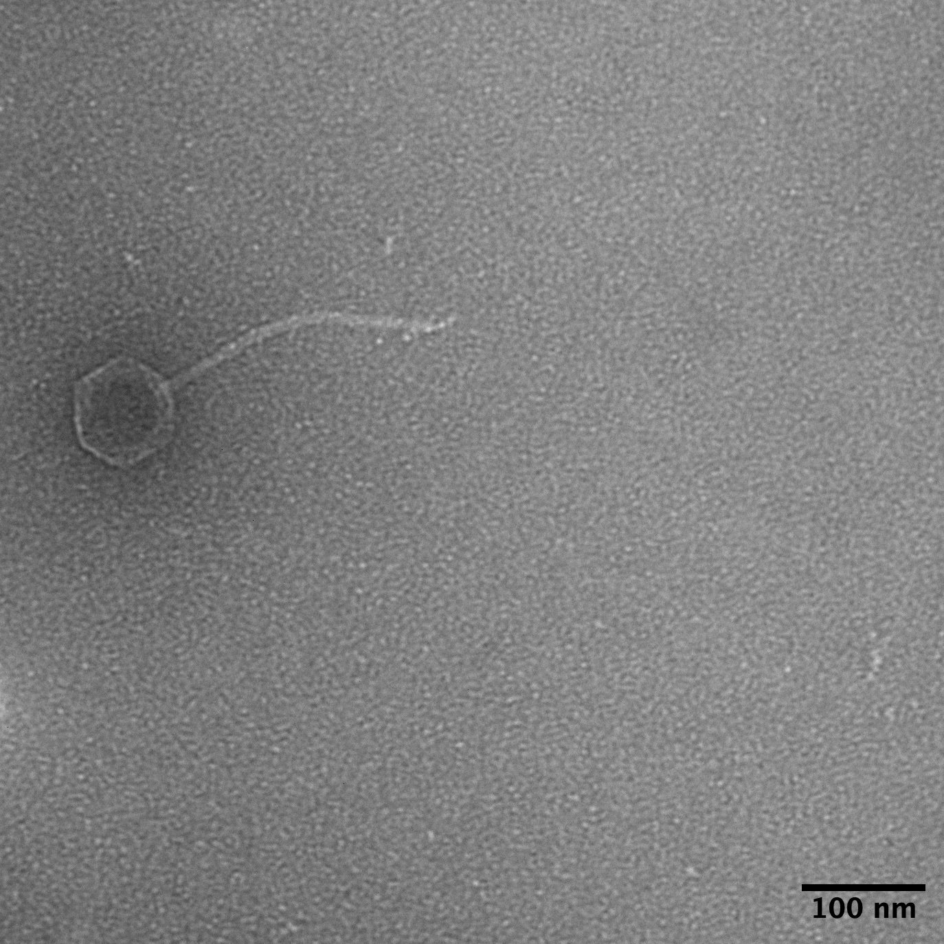
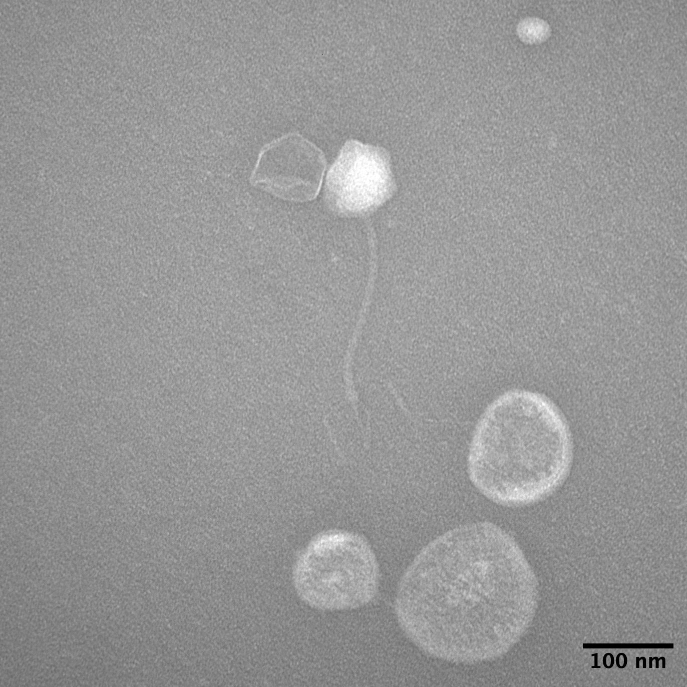
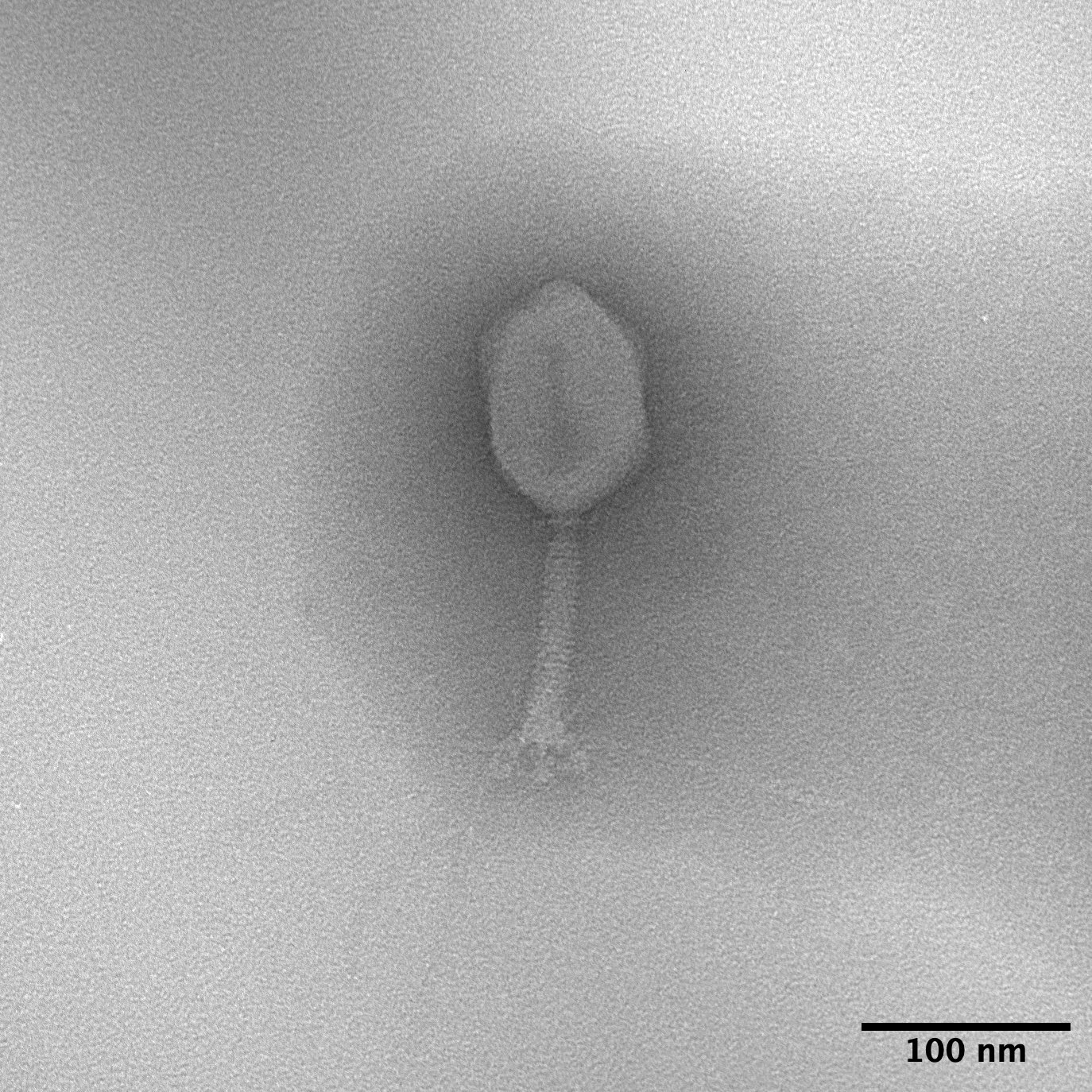
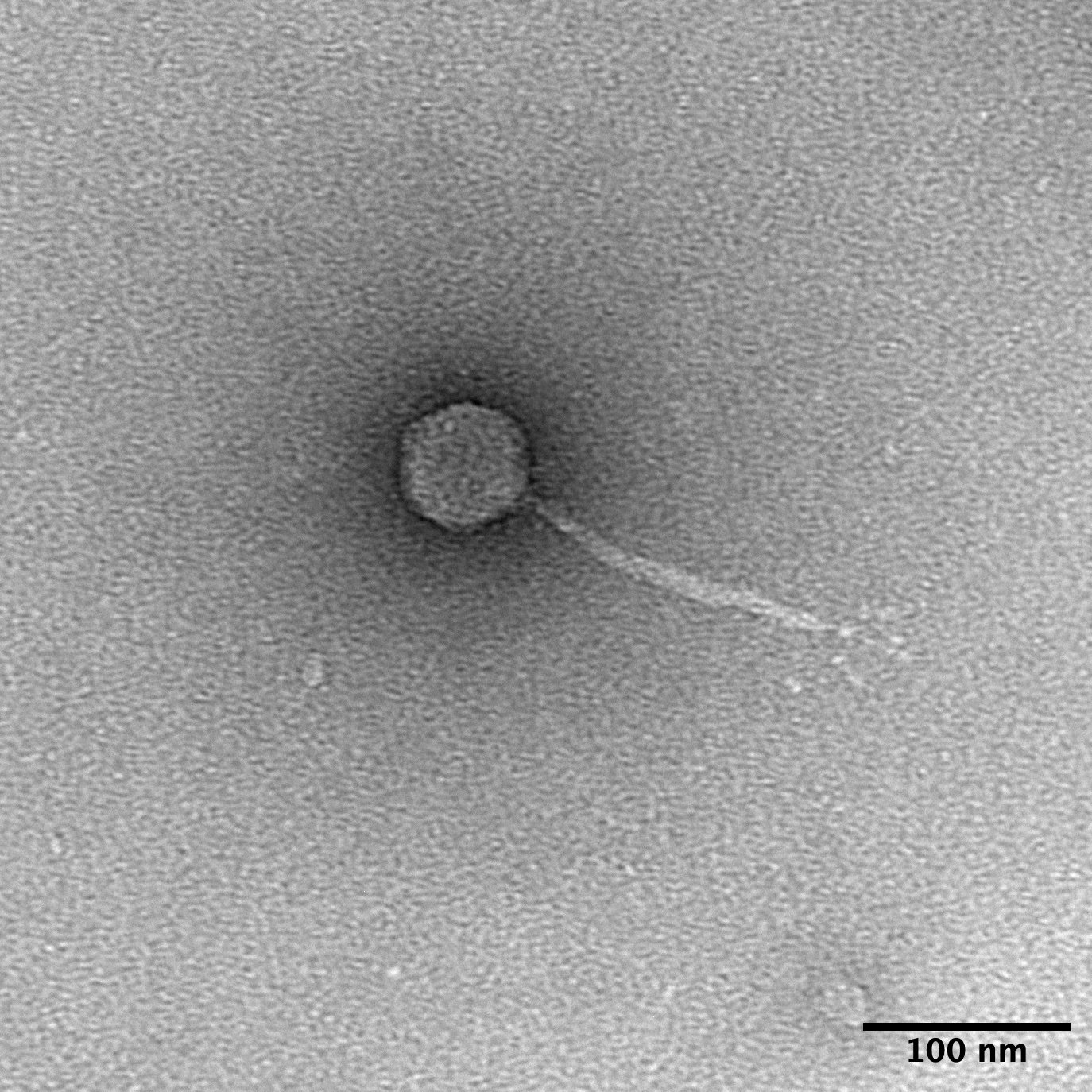
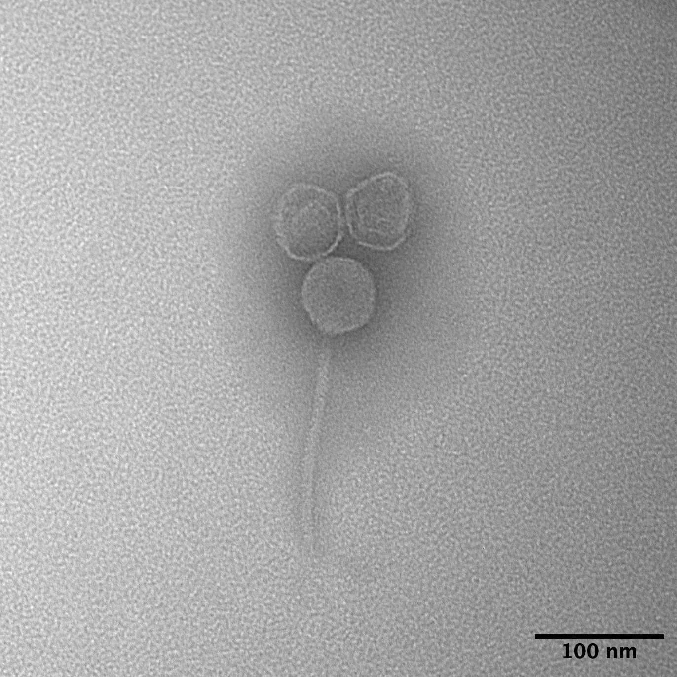
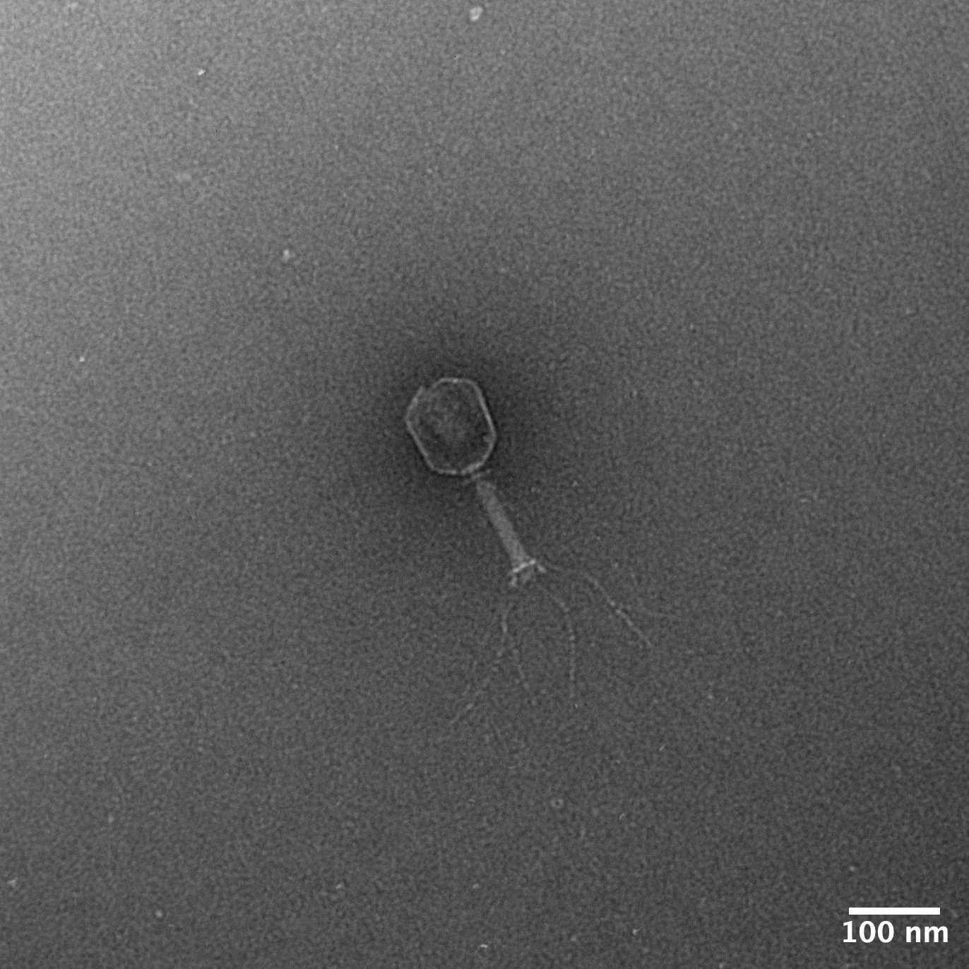
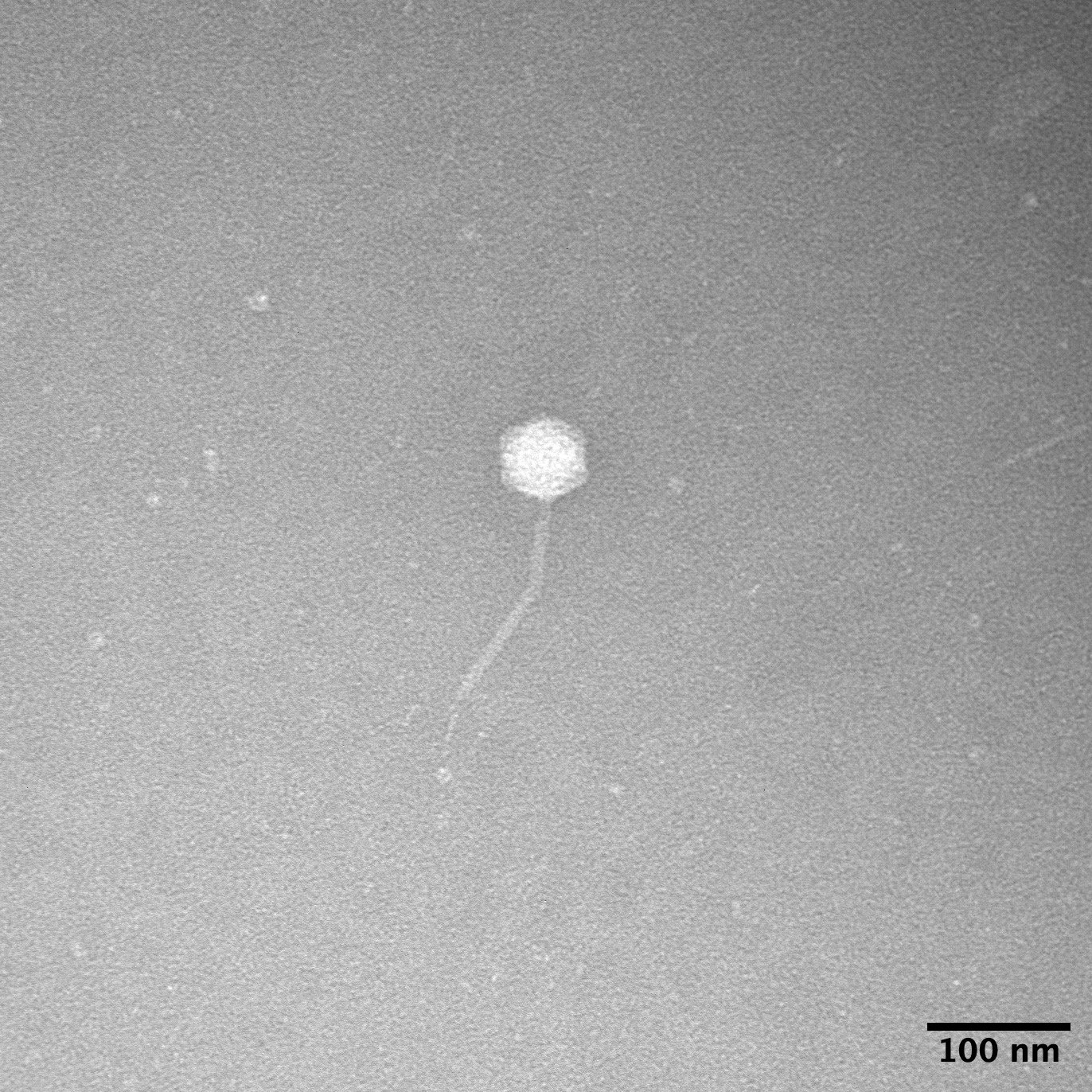
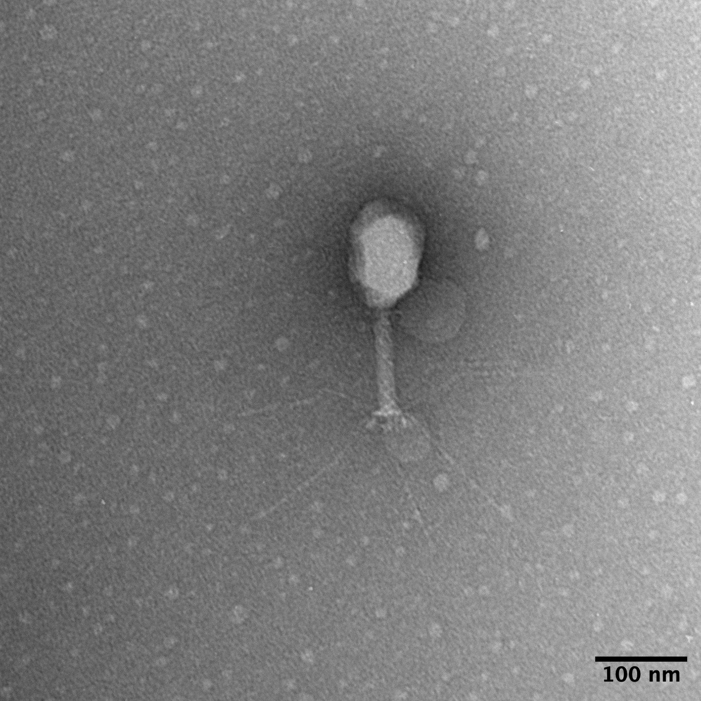
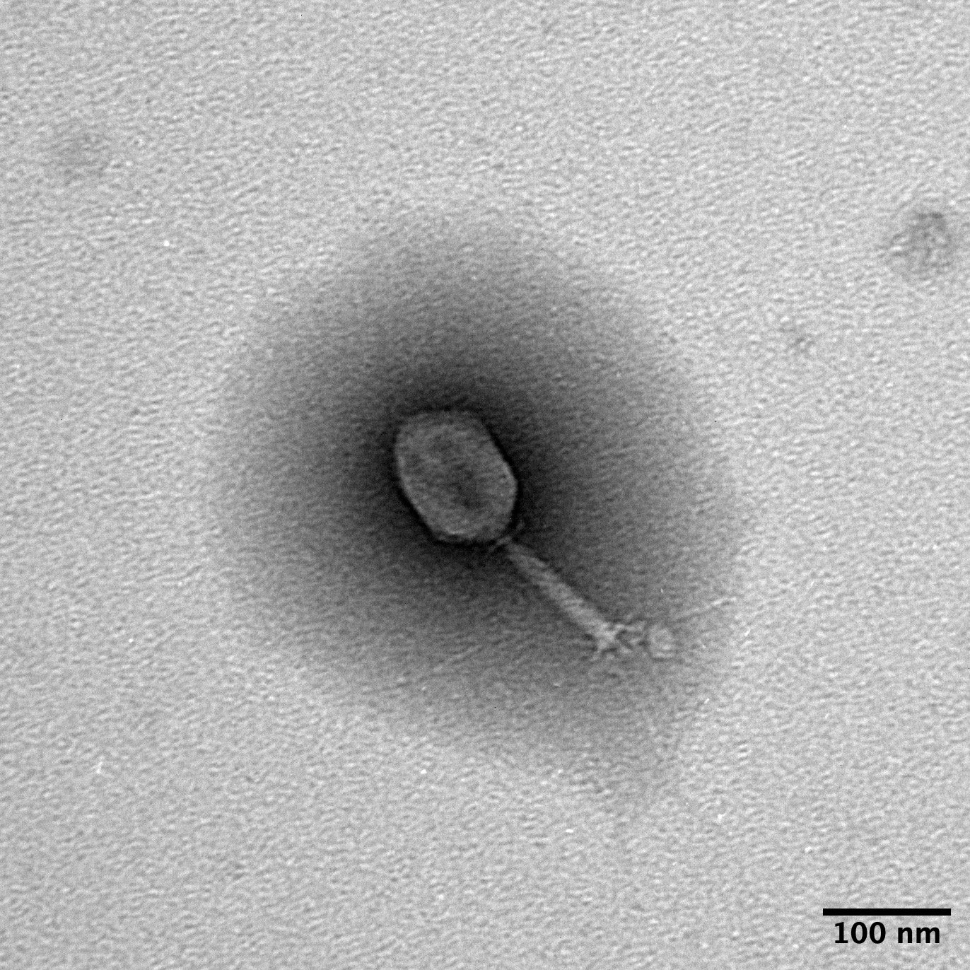
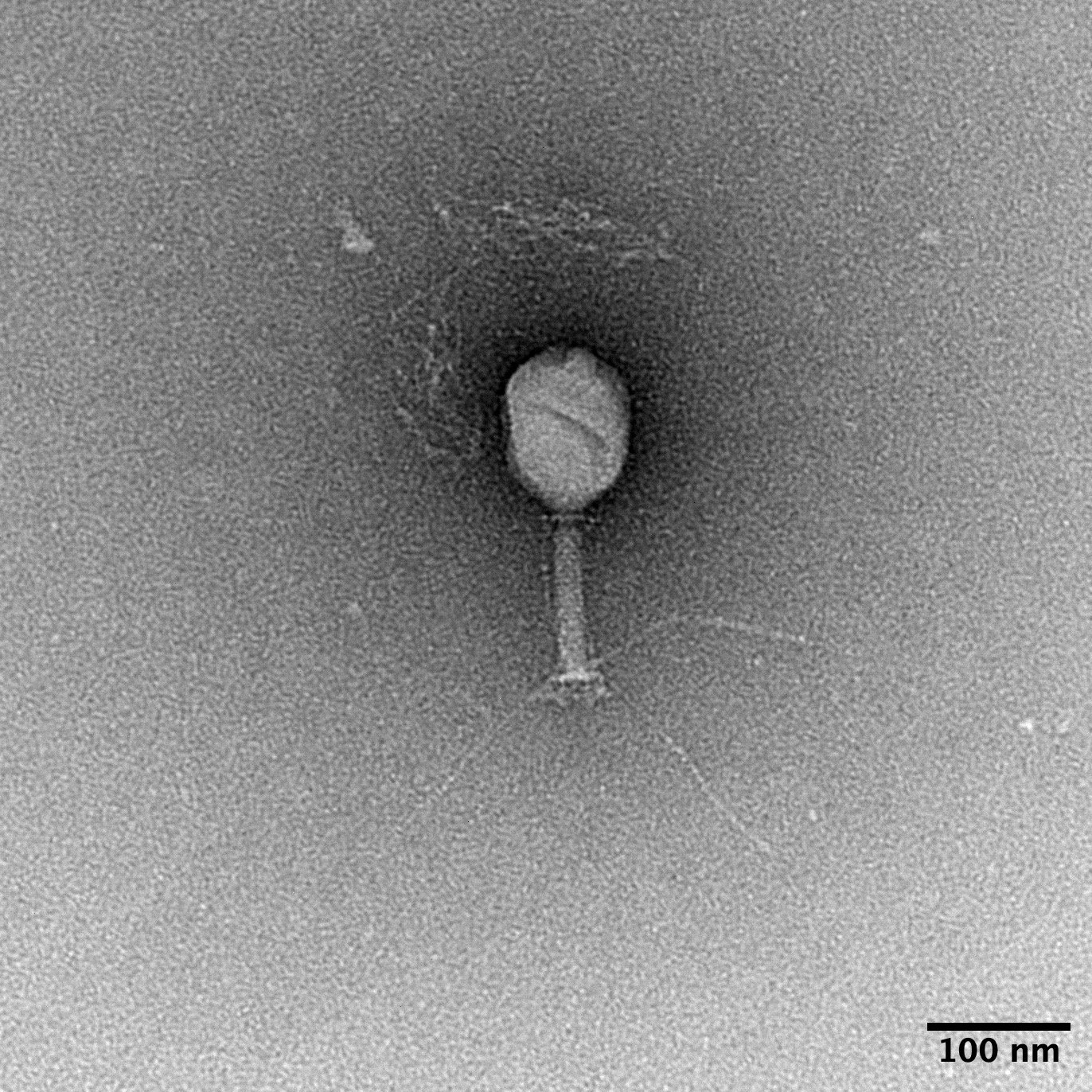
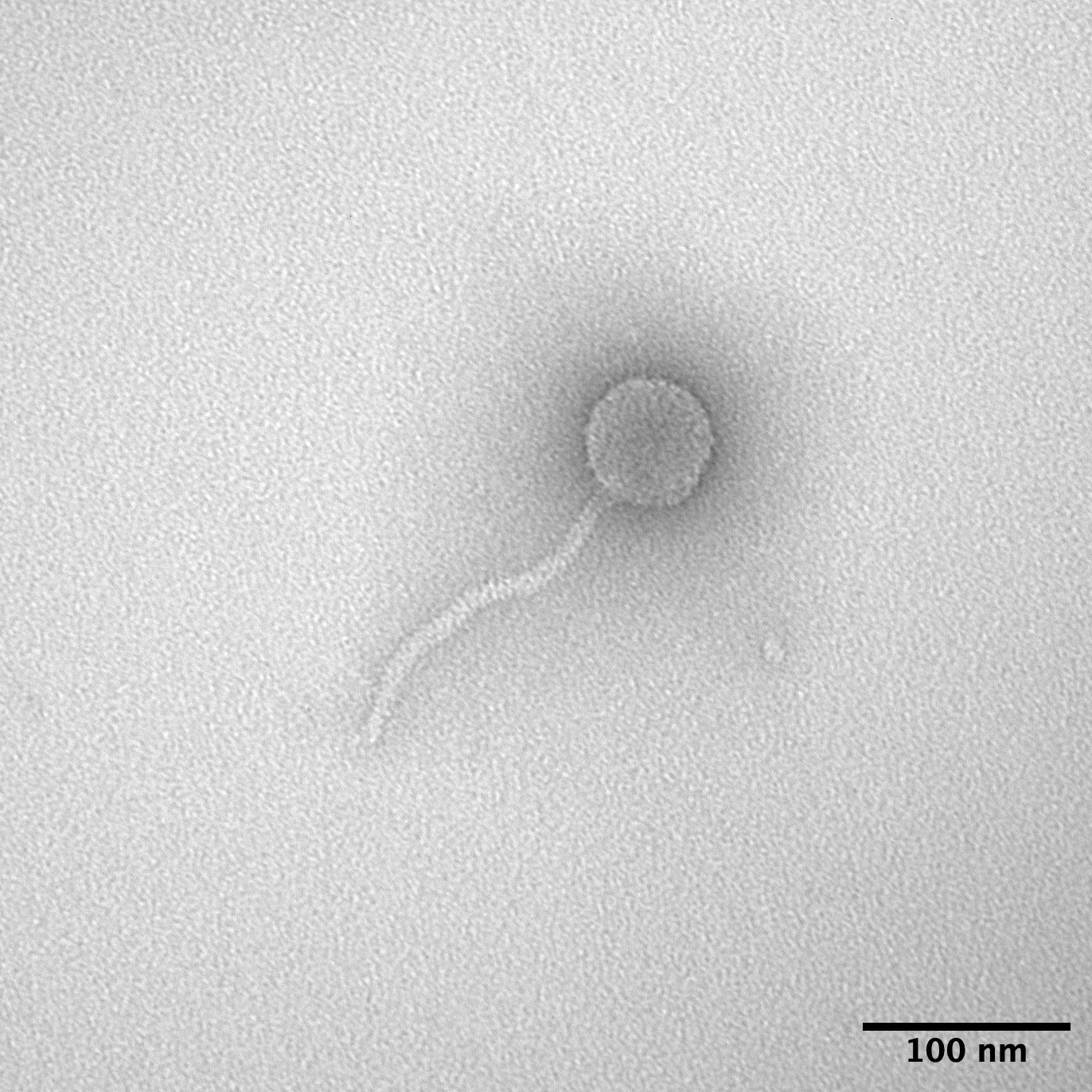
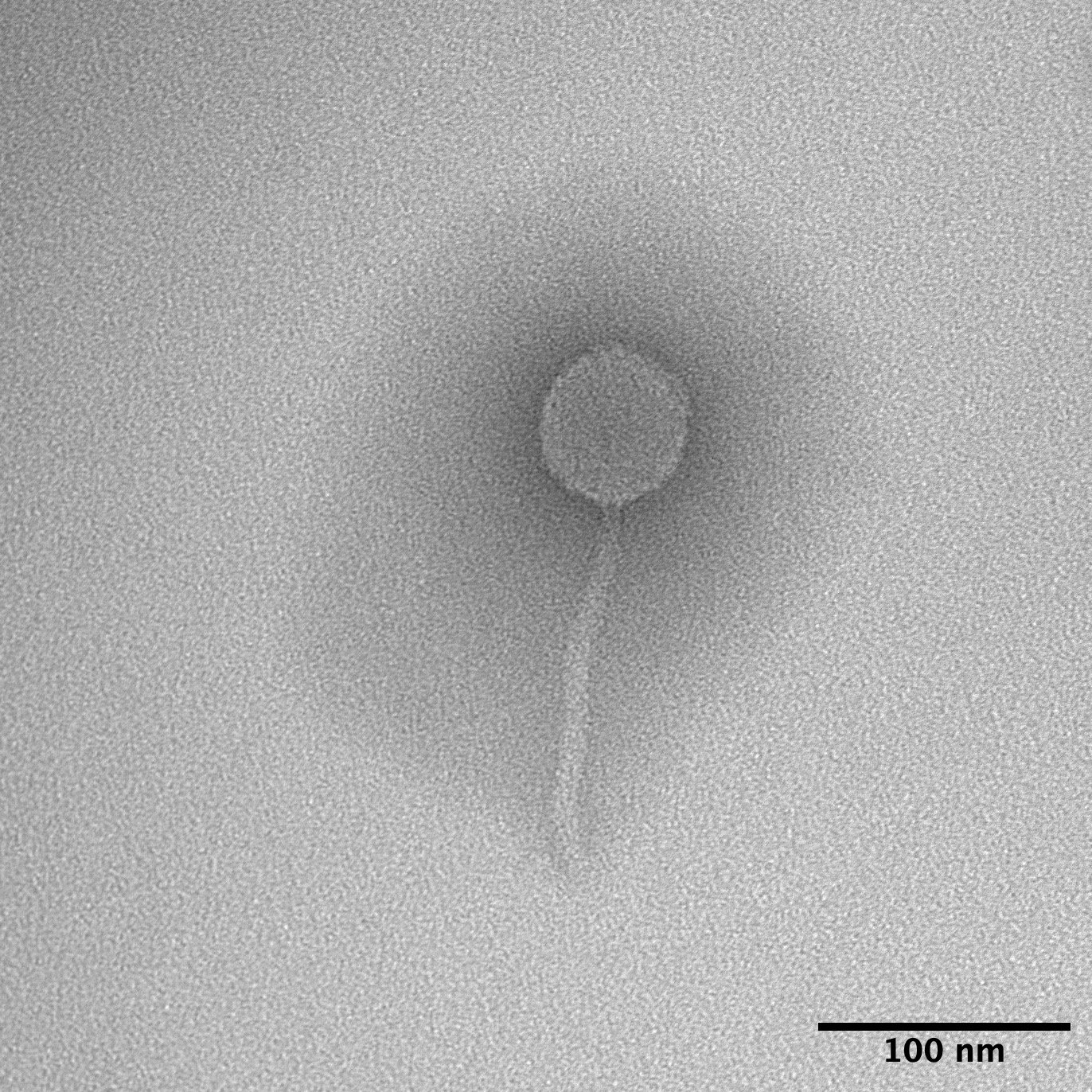
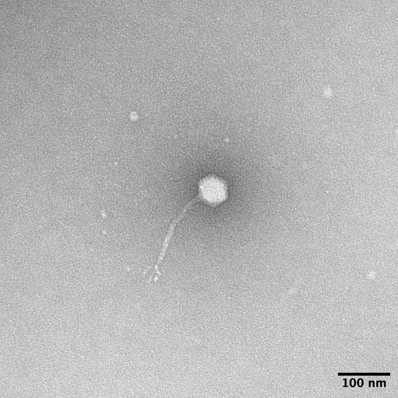
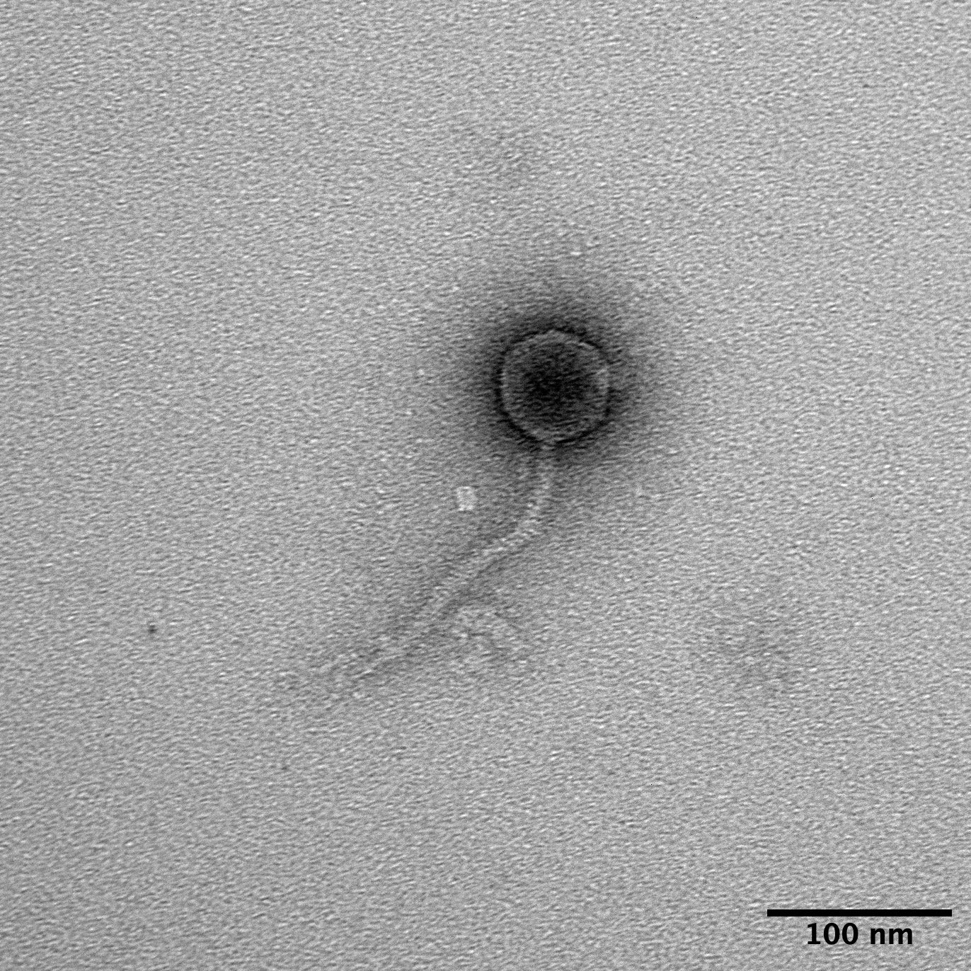
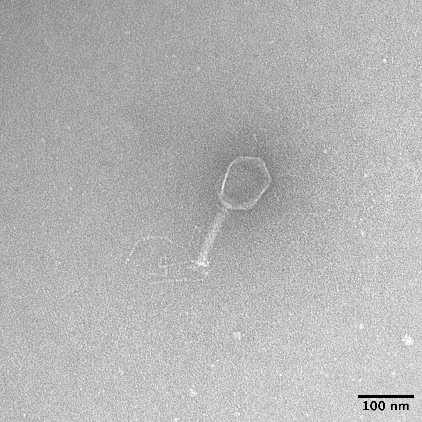
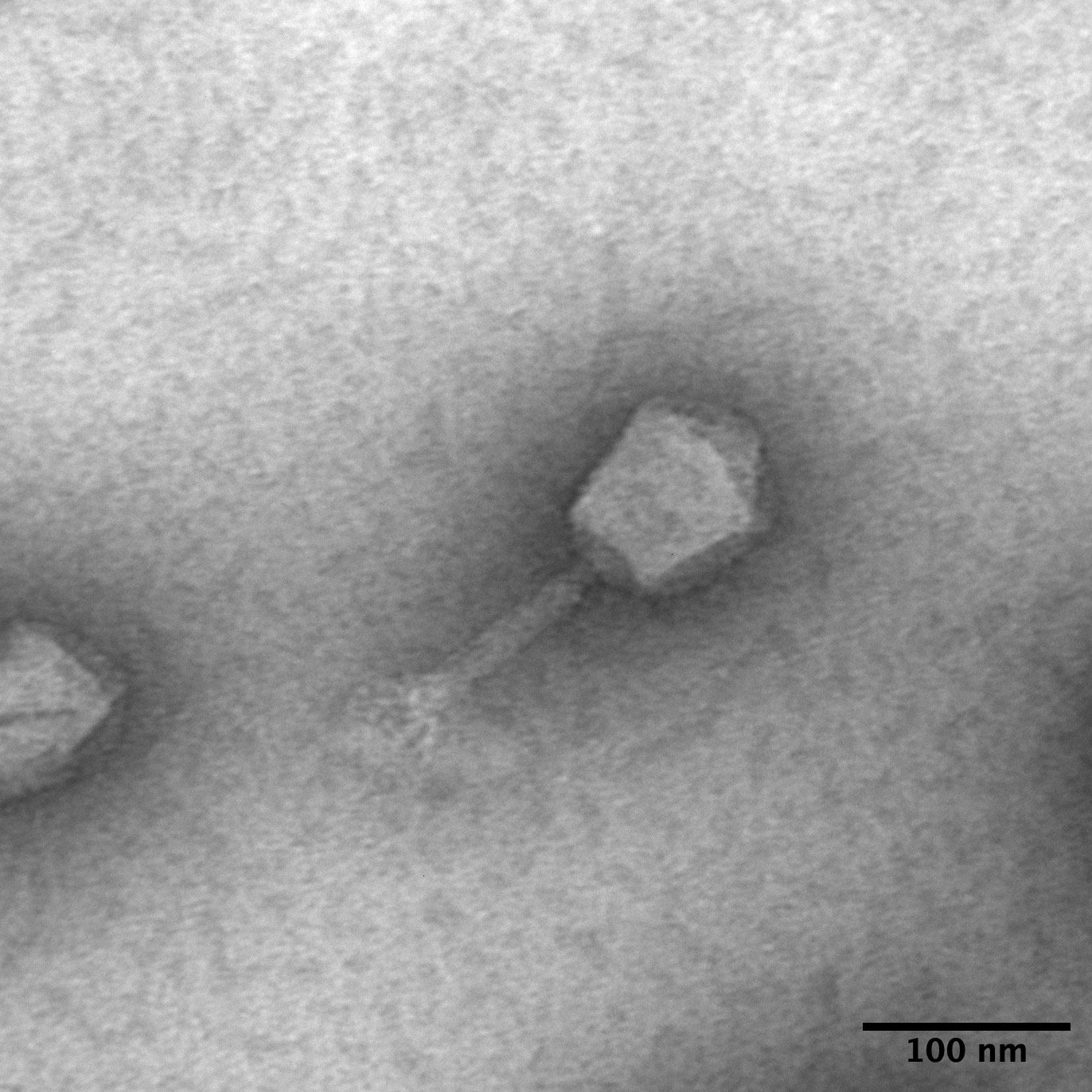
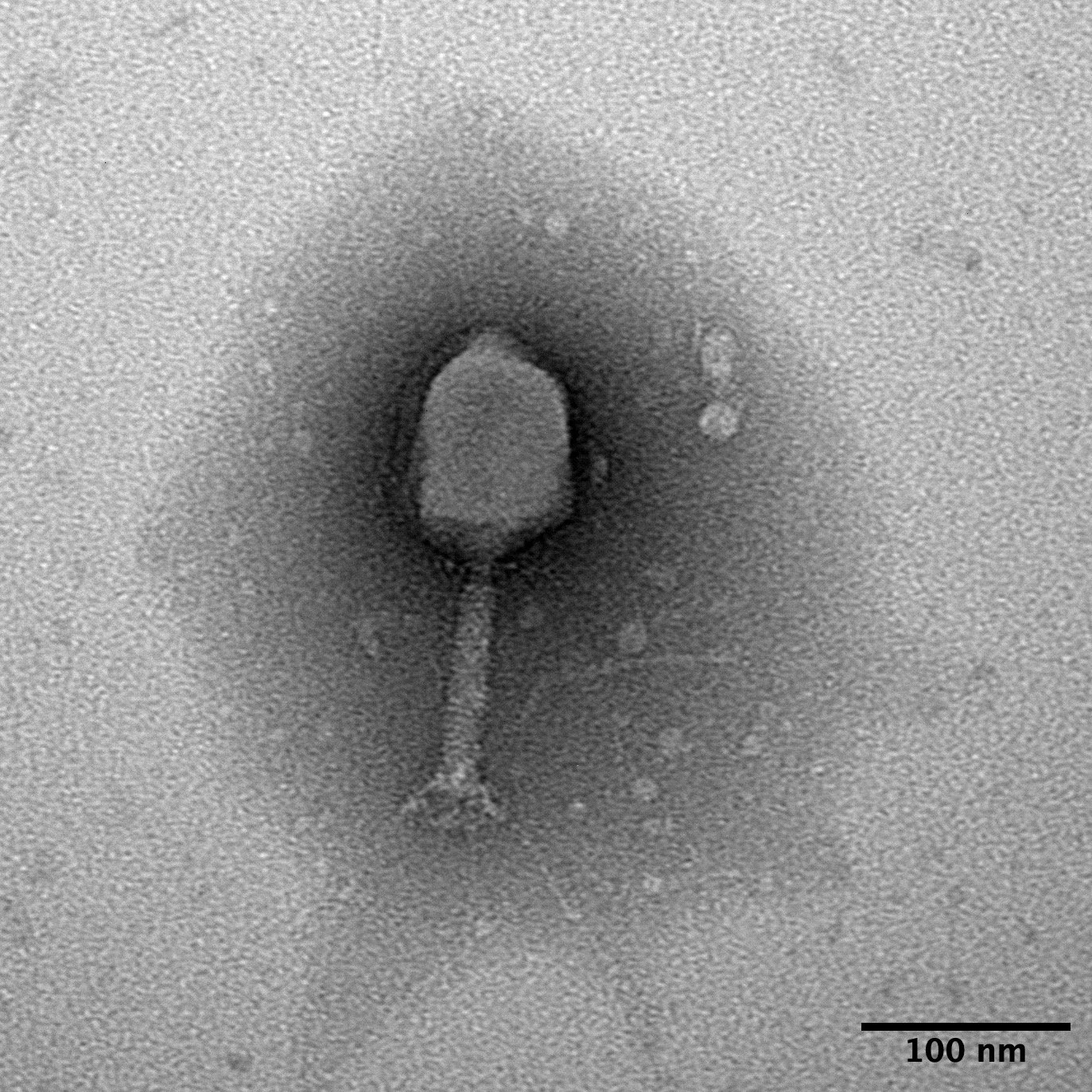
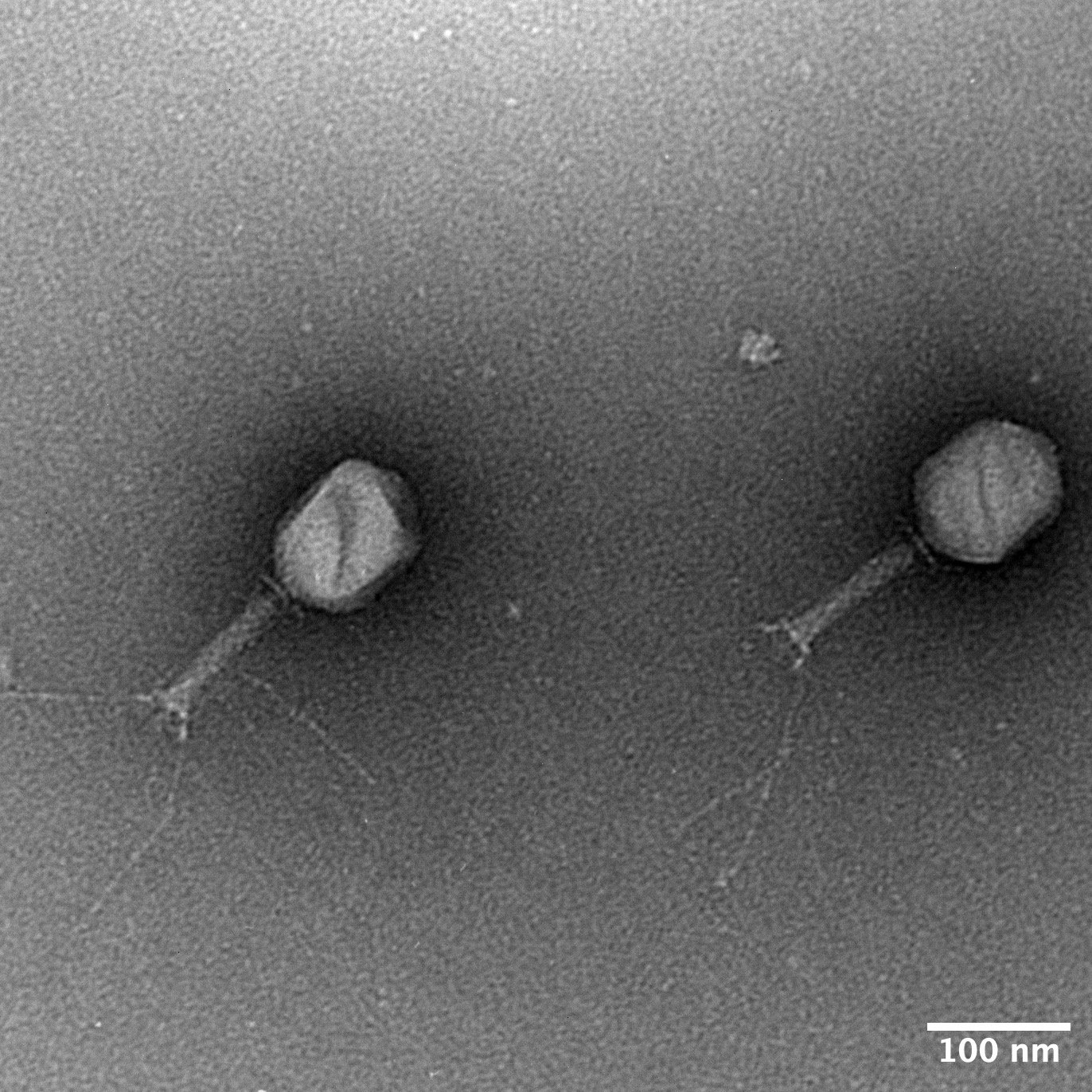
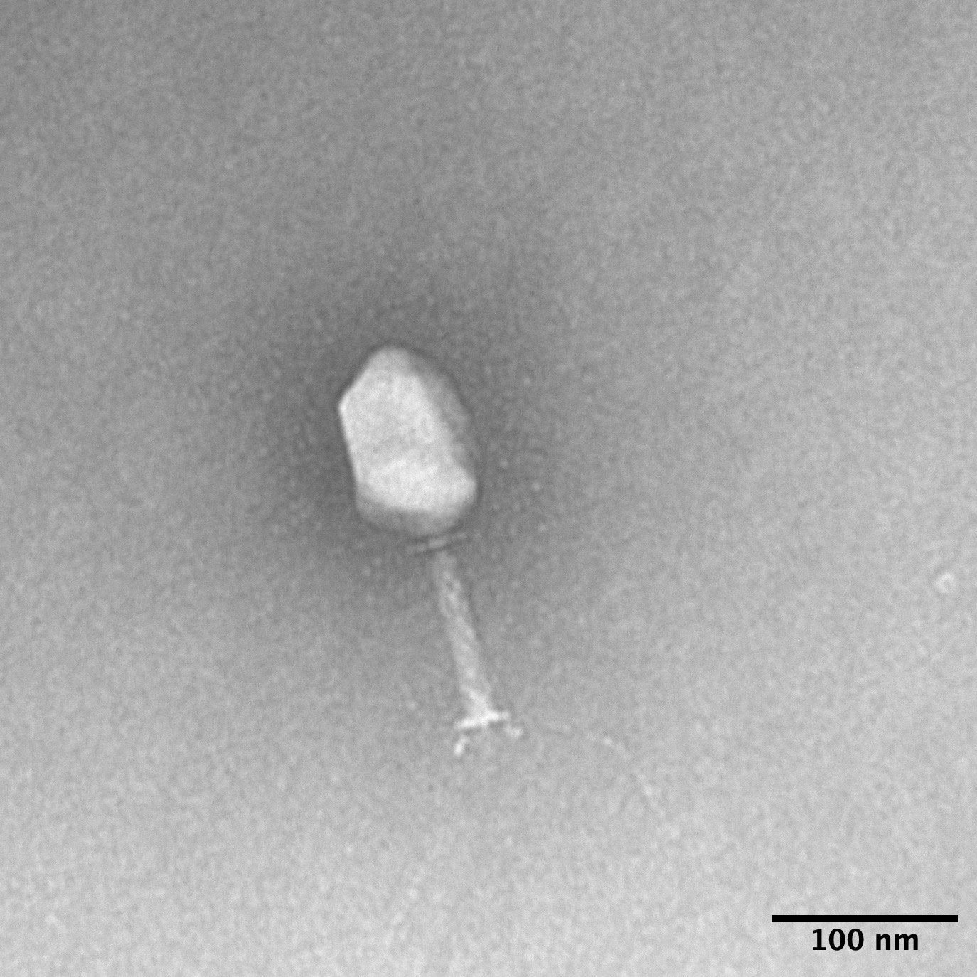
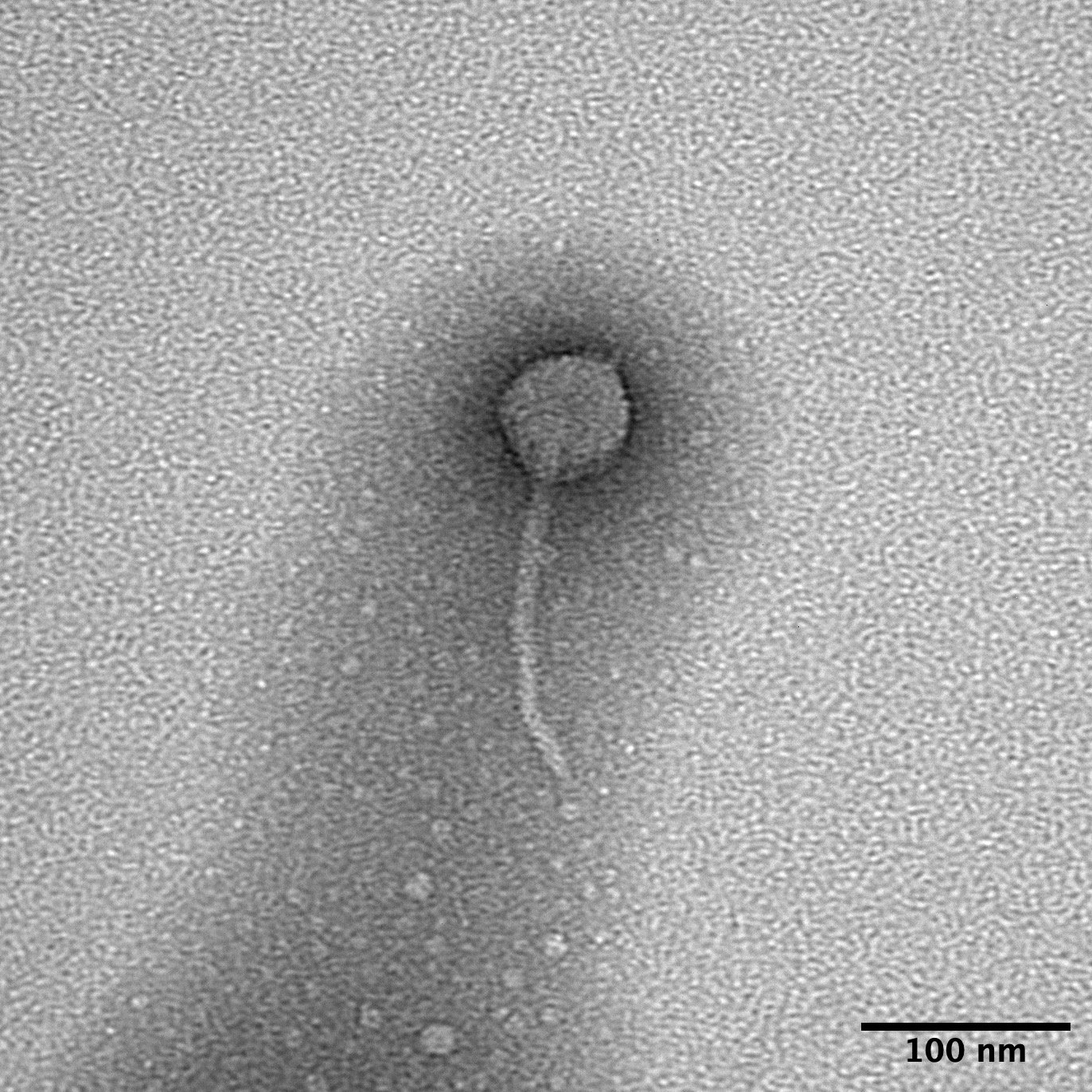
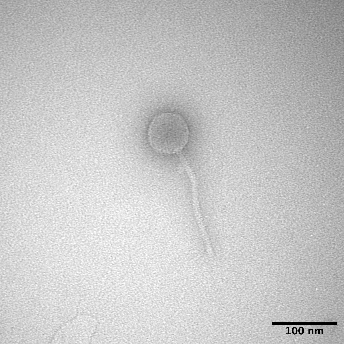
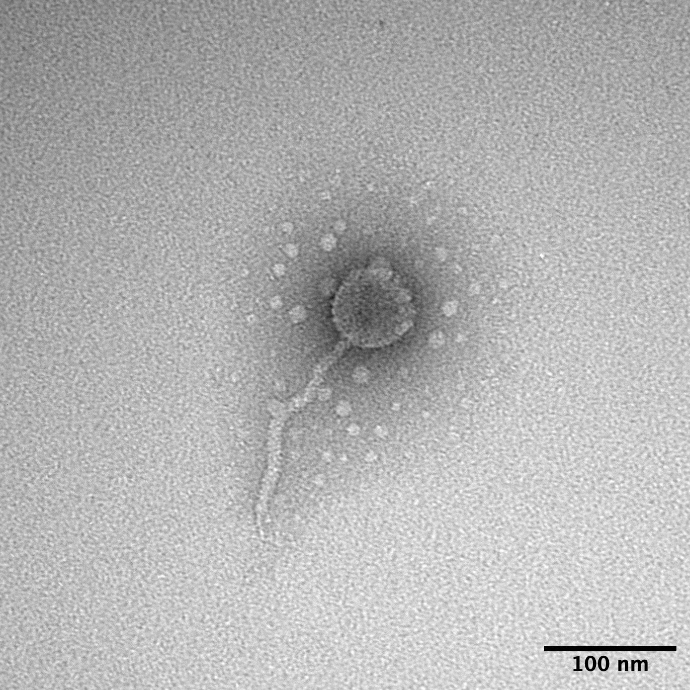
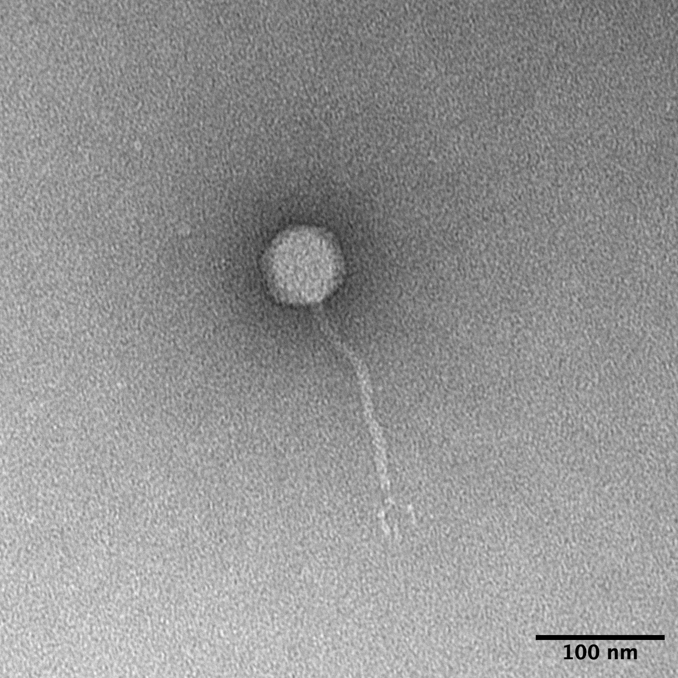
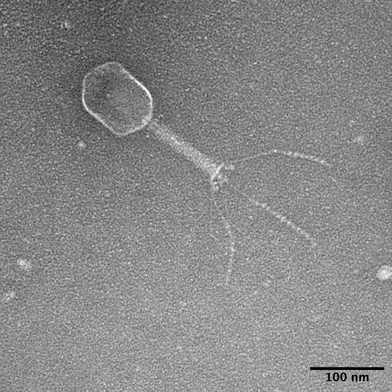
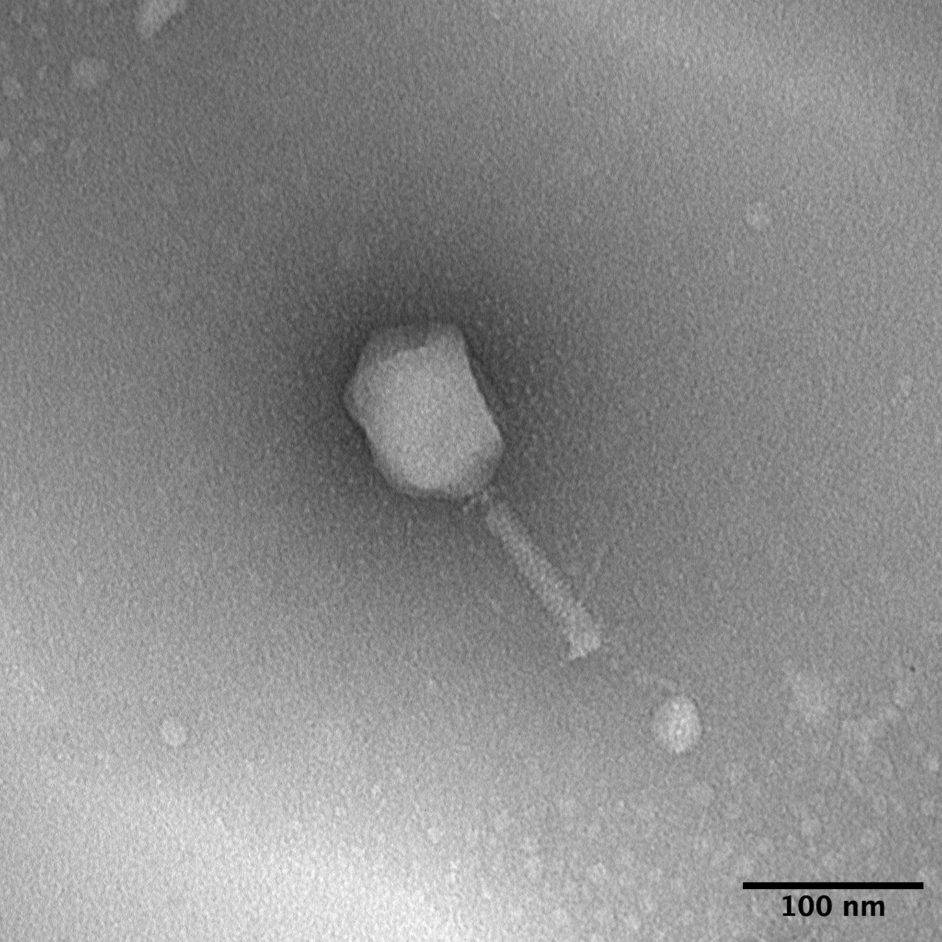
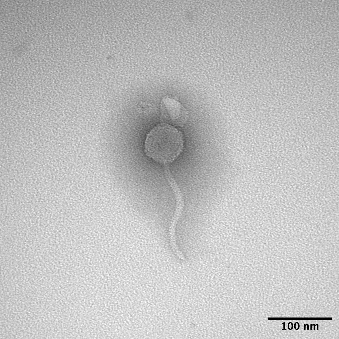
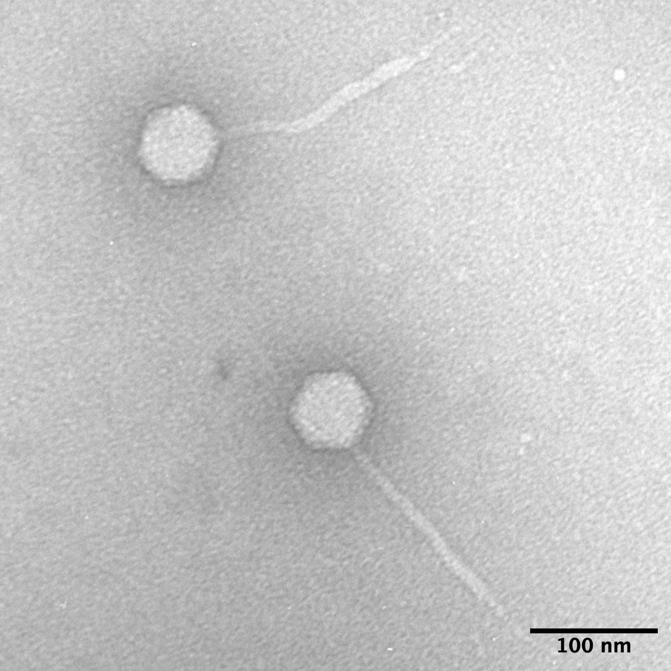
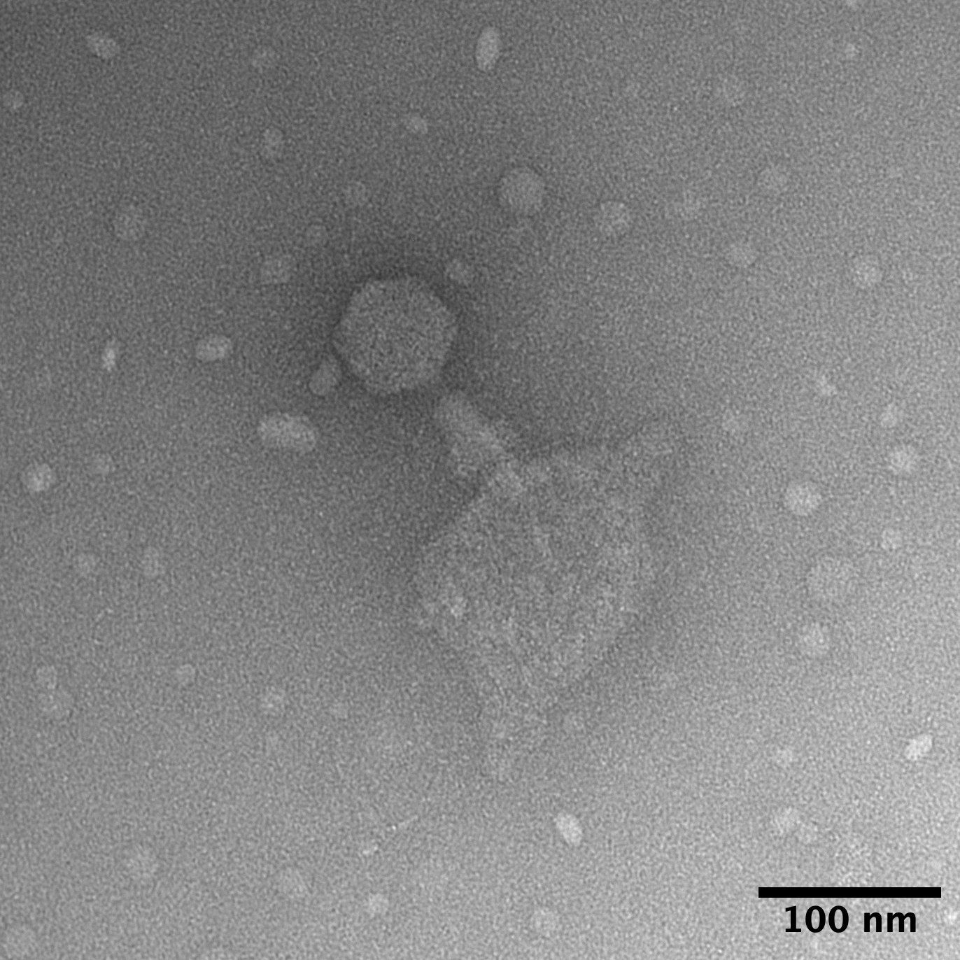
See the Transmission Electron Microscope (TEM) images for the Klebsiella phages below
All phages were imaged on glow-discharged carbon-coated type-B 400 mesh grids (Ted Pella), negatively stained with 5% Ammonium Molybdate (w/v) and 0.1-1% Trehalose. Phage virions were visualized and captured on a transmission electron microscope (TEM FEI Tecnai T12) at an acceleration voltage of 80 or 120 kV and examined at 16,500–105,000 x magnification. Fiji software v 2.9.0 was used to measure the phage lengths and to crop the images to scale. Scale bar is for 100 nm.
Roth01
Roth04

Roth08

Roth09
Roth10

Roth16
Roth17
Roth19
Roth20
Roth21

Roth22

Roth23

Roth24
Roth26
Roth27

Roth30

Roth32

Roth34
Roth37
Roth39
Roth41
Roth42
Roth44

Roth47

Roth49

Roth50
Roth51
Roth61
Roth66
Roth67
Roth68
Roth71
Roth72
Roth74
Roth75
Roth76
Roth77
Roth78
Roth79
Roth80
Roth83
Roth84
Roth85
Roth87
Roth88
Roth90
Roth93
Roth96
RothC

RothD

RothG

RothJ

RothI